

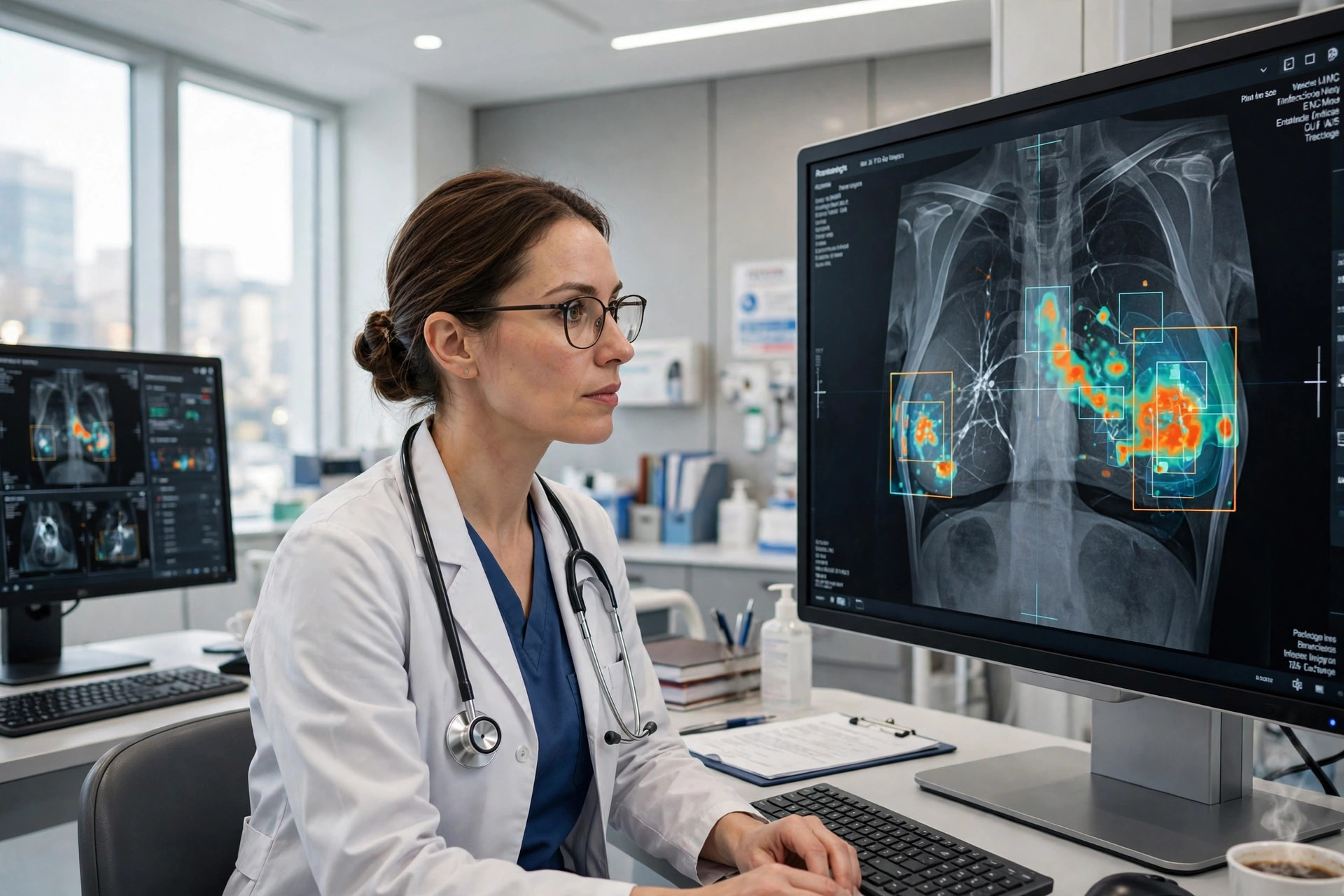
Breast cancer is most treatable when it is found early, yet the screening process designed to catch it still depends heavily on human eyes working through large numbers of detailed images. A small tumour can be easy to miss, dense tissue can hide warning signs, and a normal-looking scan can occasionally conceal a cancer that becomes obvious only months later. Across the United States and Europe, technology — and artificial intelligence in particular — is now changing that process, helping clinicians spot disease sooner and reduce the number of cancers that slip through.
The shift does not replace radiologists. Instead, new tools act as an additional reader, scanning mammograms and other images for patterns that deserve a closer look, prioritising the cases most likely to need attention and giving doctors more information before they decide whether a patient should be recalled, biopsied or reassured.
An Extra Set of Eyes on Every Scan
Traditional mammography screening often involves one or two radiologists independently reviewing each image. AI-assisted systems add a further layer of analysis. Trained on large collections of previous scans and outcomes, the software can highlight subtle asymmetries, tiny calcifications and changes from a patient’s earlier images that might otherwise be overlooked during a busy clinic.
That assistance matters because screening is a volume business. Radiologists may review dozens of cases in a session, most of them normal, while searching for the few that are not. Fatigue and distraction are human realities. An algorithm does not tire, and it applies the same scrutiny to the last scan of the day as to the first. Clinicians say the best systems function less like a replacement expert and more like a tireless colleague who quietly points and says: look here again.
The trend reflects a broader movement in medicine toward intelligent devices, similar to the consumer shift documented in coverage of how AI gadgets are transforming everyday life, but with far higher stakes and far stricter demands for evidence.
What the Evidence Is Showing
Large trials are beginning to quantify the benefit. Reporting by CBS News on technology and breast cancer detection has highlighted how hospitals are using AI-assisted imaging and other new tools to catch tumours earlier in routine care. In Sweden, the MASAI trial, which involved more than 105,000 women, found that AI-supported mammography increased detection and was associated with fewer interval cancers — cancers diagnosed between scheduled screenings — without increasing false positives. Follow-up reporting summarised by Medical News Today on AI-assisted mammography noted fewer invasive and aggressive cancers in the AI-supported group, alongside a substantial reduction in radiologists’ reading workload.
Interval cancers matter disproportionately because they tend to be faster-growing and are often diagnosed later, when treatment is more intensive. Reducing them, even modestly, could translate into simpler treatment and better survival for many patients. Earlier detection can also mean smaller tumours, less extensive surgery and, in some cases, avoiding more aggressive therapy altogether.
Technology is improving other parts of the pathway too. Three-dimensional mammography, known as digital breast tomosynthesis, creates layered views that help separate overlapping tissue. Contrast-enhanced imaging and advanced ultrasound can clarify uncertain findings, while AI tools for pathology and risk prediction are being tested to estimate which patients need closer surveillance in the first place.
Benefits, Limits and Unanswered Questions
For patients, the promise is practical: fewer missed cancers, fewer anxious recalls for findings that turn out to be harmless, and faster answers. For health systems facing shortages of specialist radiologists, AI triage can also help direct scarce expertise to the cases that need it most, rather than spending equal time on scans that are almost certainly normal.
Doctors and researchers are careful about the limits, however. Algorithms are only as good as the data they were trained on, and performance must be checked across different ages, ethnicities, breast densities and imaging equipment. A tool that works well in one screening programme may need adjustment in another. Oversight, transparency and clear accountability remain essential: when an AI system and a clinician disagree, there must be a defined process for resolving that disagreement, and final responsibility still rests with the medical team.
Access is another concern. New imaging technology is expensive, and there is a risk that its benefits concentrate in well-funded urban centres while rural or under-insured patients are left with older methods. Advocates argue that if AI can reduce workload and prevent later-stage disease, it could eventually lower costs as well as improve outcomes — but that will depend on deliberate policy choices, not technology alone.
A Screening Future That Arrives Earlier
The direction of travel is nevertheless clear. Screening is moving from a single snapshot, read once, toward a continuous, data-rich process in which prior images, personal risk factors and algorithmic analysis all inform the decision. Some researchers are even testing whether AI scores from scans taken years earlier can flag patients who merit closer monitoring long before a tumour is visible.
For the millions of women who attend screening each year, that future would feel reassuringly familiar — the same appointment, the same machine — with one important difference: behind the scenes, a second, digital reader would be working to make sure that fewer cancers are missed, and that those which are present are found at the moment when treatment can do the most good.
